It was such a fine evening as the sun rays cast a golden glow onto the trees. I had completed my day's work and looked forward to relaxation. I thought I would just drop into a Veterinary Surgery of my old friend to see what type of spay hook my peer had bought from Australia. His Veterinary Technician had said they were excellent hooks.
There are many makes of spay hooks and I am always on the look out for the best type which is well designed and easy to use. As if there are such instruments but one never knows.
 "Do you want to see a bladder
stone surgery?" the Vet Technician whom I knew for more than 10
years asked me as I was leafing through the various pages of skin
diseases in dogs in the several text books at this Surgery. A
veterinarian on duty was performing this surgery. I really wanted
to enjoy this beautiful sunset and the fresh air. "Well, I could
just see a while and go off," I thought this was a run-of-the-mill
urolith case.
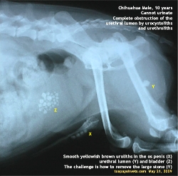
 However, this case was an exceptionally challenging case due to the fact that there were 3 places where uroliths had formed or got stuck.
From my view of the X-ray, the main problem was the urolith (Y) stuck at the bend of the urethra, between the bladder and the penis. The stones at X and Z could be removed by cystostomy (cutting open the bladder) and urethrostomy (cutting open the urethra behind the os penis). But God had placed one large stone which cannot be removed by cutting as the stone was inside the urethral lumen inaccessible to surgical blades.
How to get this stone at Y out? There would be 2 ways:
1. Use a rigid catheter to push the stone into the bladder. The stone (Y) would then be removed via the bladder incision. However there would be traumatic damage to the urethra tissues as the rigid catheter forces its way trying to dislodge the large stone (Y). This result in profuse of bleeding and that will not be good for the dog. The success, in my opinion, is not be guaranteed. This large stone was the cause of urinary obstruction. The bladder had distended to over 30X its normal size and was going to rupture in this case.
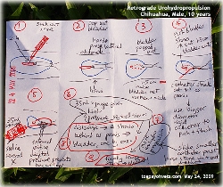
2. Retrograde urohydropropulsion. What this method means is to flush normal saline via the penile orifice into the urethra to flush out the stone at Y into the bladder. In theory, this sounded easy. In practice, this could be hellish.
Procedure:
1. General Anaesthesia. The 10-year-old male Chihuahua was put on an IV drip. It weighed 2.5 kg. Injectable anaesthesia was used. 0.2 ml of 20% xylazine and 0.05 ml of ketamine IV was given. The dog went into general anaesthesia. Topping up with small amounts of Zoletil 50 IV given via the IV drip at 0.1 ml was used when necessary.
2. Surgery.
2.1 Ventral recumbency. The bladder had distended to near the umbilical area. A 4-cm midline incision cranial to the penile orifice was made. The bladder was exposed.
2.2 Cystocentesis. A 21-G needle with a 10-ml syringe was inserted midway between the vortex and neck of the bladder, on the ventral side. Over 100 ml urine was extracted. The bladder was now half full.
2.3. The assistant used his two hands to press inwards on the lower abdomen, ventrally and from laterally. The bladder popped out of the skin incision.
2.4 Cystotomy. The bladder was incised 1.5 cm where the needle was inserted. The stones were removed from the bladder using forceps and flushing. This was the easy part of surgery.
2.5 Retrograde urohydropropulsion. A 10-ml syringe with normal saline would not be effective as the pressure would be too low. A 30-ml syringe was used. The rigid catheter (not flexible one) was used. It got stuck around 10 cm after passage as there were stones behind the os penis.
 The
penile orifice was closed by the thumb and forefinger of the
assistant while the saline was forced in. This was repeated a few
times. Still there was no stone or saline coming out from the
bladder. There were 4 stones behind the os penis and these were
big stones.
The
penile orifice was closed by the thumb and forefinger of the
assistant while the saline was forced in. This was repeated a few
times. Still there was no stone or saline coming out from the
bladder. There were 4 stones behind the os penis and these were
big stones."Remove the stones behind the os penis by cutting open the urethra (urethrostomy) behind the os penis" the assistant recommended. There seemed to be no headway. I was the observer and therefore needed not sweat this out. So, should urethrostomy behind the os penis to remove the 4 stones easily be done?
As an observer, it was easy for me to comment: "Even if you remove the 4 stones by cutting the urethra behind the os penis, you still cannot remove the large stone (Y) as it is deep inside the urethra. You can use the rigid catheter to push it out but there will be a lot of bleeding. Try flushing again. How about rectal pressure?"
Rectal pressure meant putting a finger inside the rectum and then onto the urethra. This would help to increase the pressure of the saline to flush out the stone (Y) when the finger is released after another 30 ml of saline was injected via the catheter. Now, this method was not practical as the dog was a small breed and in any case, it was on ventral recumbency.
Retrograde urohydropropulsion was tried several times again. The saline came out via the penile orifice each time the assistant tried. He used a bigger diameter catheter to push the 4 stones. There seemed to be some progress.
"How about massaging the urethra behind the os penis?" I suggested. As an observer, I was not so much stressed out as this was not my case. So I could make such a suggestion. The Vet Technician massaged the urethra with his fingers. Up and down. Then he resumed flushing.
I don't know whether my suggestion really worked or not. One stone got flushed out of the urethra into the bladder and was picked out. More urohydropropulsion. 3 more stones came out. There were of similar sizes. "These are from behind the os penis," the Vet stated as their sizes were similar to those seen on the X-ray. "There is now the big stone still stuck."
By this time, the smaller rigid catheter was used and it went further into the urethra and appeared out in the bladder incision. The team was elated but silently happy. We still had this one last "diamond" inside. If only it was a real diamond. What should be done now?
As an observer, I could advise: "Push the catheter in and out of the bladder to roll the large stone loose and out of the bend of the urethra." The Vet did that a few times. Then he commenced urohydropropulsion. The smaller rigid catheter could move in and out of the bladder but not stone was seen.
Was there any hope? The Vet flushed normal saline from the opening at the bladder neck into the urethra. "Will this method be pushing the large stone into the urethra?" I asked him. He did once only. He continued flushing saline via the penile orifice and clamping the orifice shut with his thumb and forefinger to increase the pressure.
In what seemed like an
eternity, the large stone rolled out into the bladder. The stress
was over for the team. And for me too. An observer could also be
stressed when things do not move on smoothly. I was happy for the
team and for the dog.
I described this case in great detail to share my observations
with veterinary students who must study urohydropropulsion - a big
word reading the procedure from dull veterinary surgery text
books. I hope this real life case may be of use to the 4th and 5th
year veterinary students who need to pass examinations.
This case was great team work. The dog went home to a very happy
owner. Instead of dying from bladder rupture and kidney failure. I
missed a fine evening but this was a most interesting case if one
was not the operating veterinary surgeon.
NOTE TO DOG OWNERS. Get your dog treated early and promptly
when he had difficulty in passing urine and not wait till it is
too late. Not every surgery has such a successful outcome as this
case. The large stone is stuck at the bend of the urethra was due
to a long time in seeking veterinary treatment.
It was an extremely difficult case to remove such a large stone as this
part of the urethra is not accessible by surgery.
In retrospective review of the case in May 28, 2009, I wonder how
the 4 stones stuck behind the os penis could be flushed out
past the large stone (Y) one by one. The large stone (Y) was
the last to be flushed out. The urethra lumen would need to
be at least the combined diameter of the smaller and large stone
at the bend of the urethra. In theory, this was NOT possible. This
seem to be an incredible story. If I had not been present, I would
have said that this is a tall tale. Out of this world. Did God
give a hand to the mere mortals and the old dog?
In real life, many things cannot be explained. Fact is stranger
than fiction? Seeing is believing and therefore I will not expect
the reader to believe this story.
NOTES TO VETERINARY STUDENTS
Rectal pressure is applied for dogs standing. The bladder
is not incised yet. Stones are flushed into the bladder using
retrograde urohydropropulsion. In such cases, the bladder must not
be fully distended. The use of rectal pressure is to increase the
pressure, using 30-ml syringe and normal saline to flush the
stones behind the os penis into the bladder. Obviously, the stones
must be smaller than in this case. As the stones get into the
bladder, a cystostomy is then done. Alternatively, the small
stones are dissolved using dietary methods and other medication
without the need for surgery.
Injectable anaesthesia. 0.2 ml of 20% xylazine and
0.05 ml of ketamine 100mg/ml IV, separate syringes, were given to the 2.5 kg dog. It
provided excellent general anaesthesia in this case for some 15
minutes but topping up with 0.1 ml Zoletil 50 IV was easy.
Topping up was to effect. See other information at:
Animal Shelter Medicine.
I use isoflurane gas anaesthesia but injectable anaesthesia is
nowadays excellent and safe for dogs too. Topping up is necessary.
Each vet has his own preferences. I hope the vet students
understand what is the meaning of retrograde urohydropropulsion
now.
 TOA
PAYOH VETS